Клетки корня под микроскопом - найдено 60 изображений
Найдено изображений: 60
Клетки корня под микроскопом14,118 Cells Microscope Stock Photos - Free & Royalty-Free Stock Photos from Dre
25 шт., набор горлышек для микроскопа Alibaba.com29,263 Microscope Cell Stock Photos - Free & Royalty-Free Stock Photos from Drea35 Stunning Examples Of Photomicrographs Mitosis, Microscopic photography, PatteApical meristem, terminal bud vector illustration Terminal bud, Microscopic photCells of a Root Under the Microscope. Stock Image - Image of epidermis, microscoCoifa (biologia) - Wikipédia, a enciclopédia livre
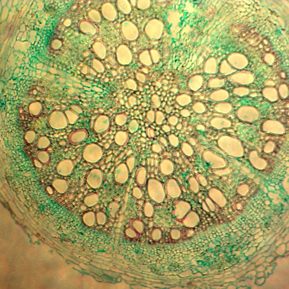
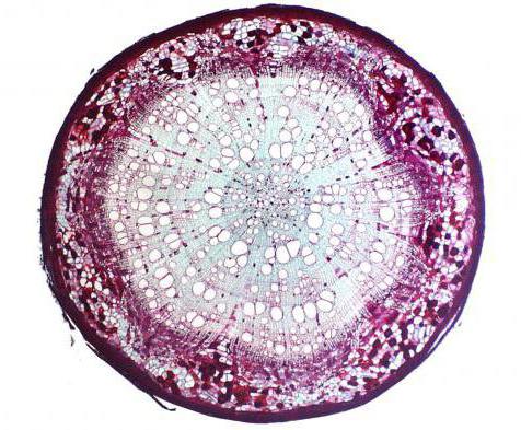
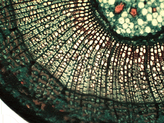
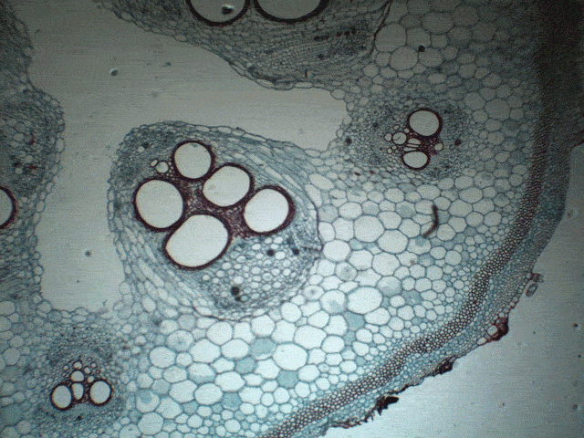
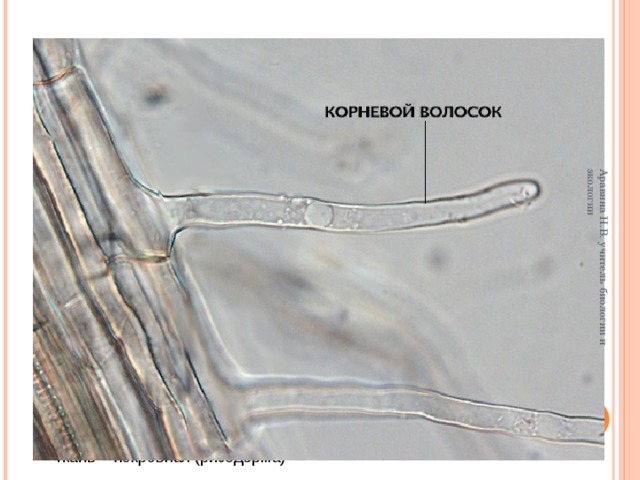
Coleus Shoot TipCross Sections of Plant Root Under Microscope View Stock Photo - Image of opticacross-section of the structure of a sugarcane root. (Debora Leite) Microscopic pDifference Between Apical Intercalary and Lateral Meristem 1 Plant cell, DiffereDisplaying 20 Images For - Metaphase Mitosis, Display, DecorIris root, light micrograph - Stock Image C002/7006 Microscopic photography, MicLearn About Plant Cell Types and How They're Like Animal Cells Plant cell, Micro
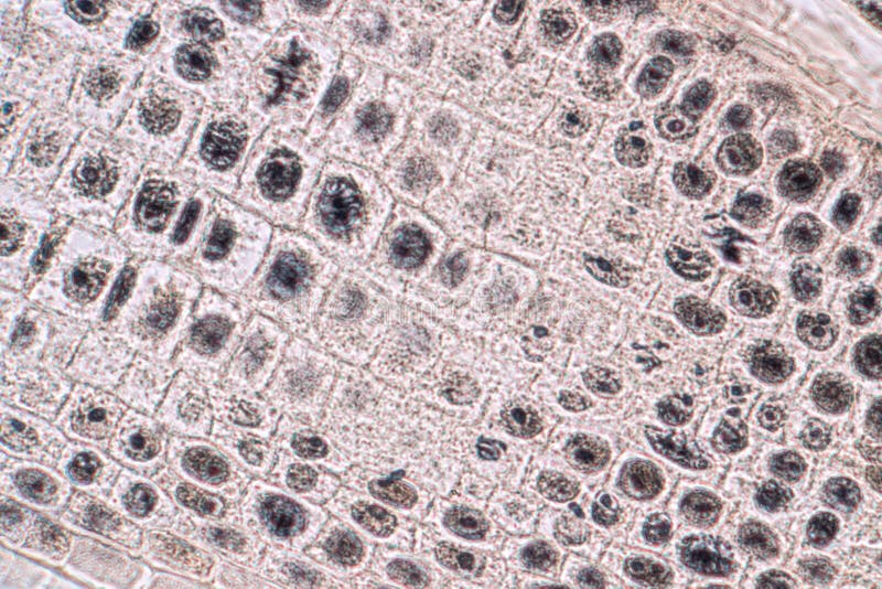
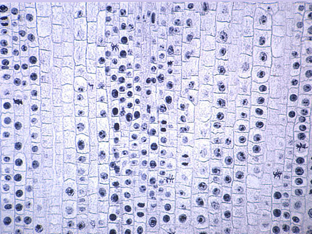
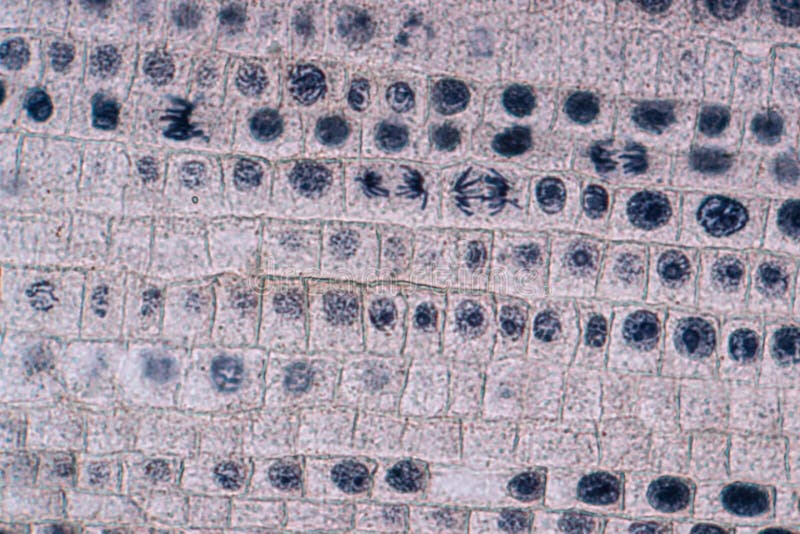
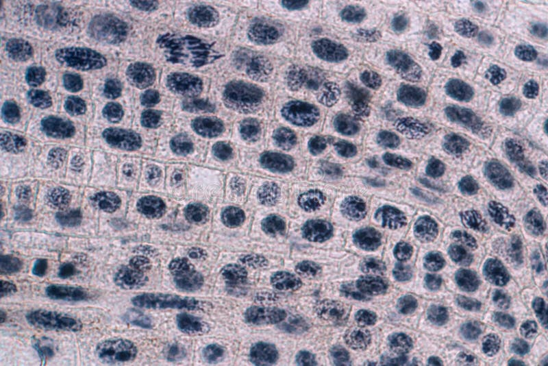
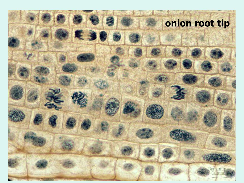
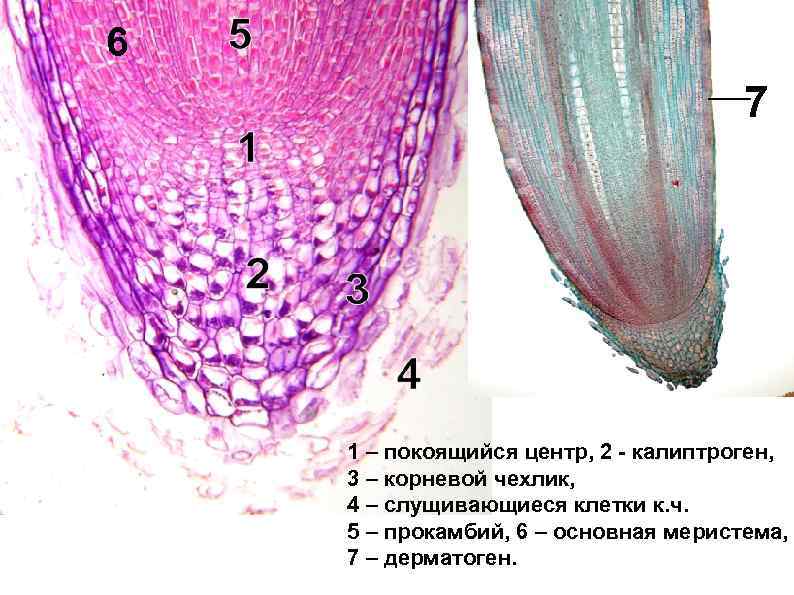
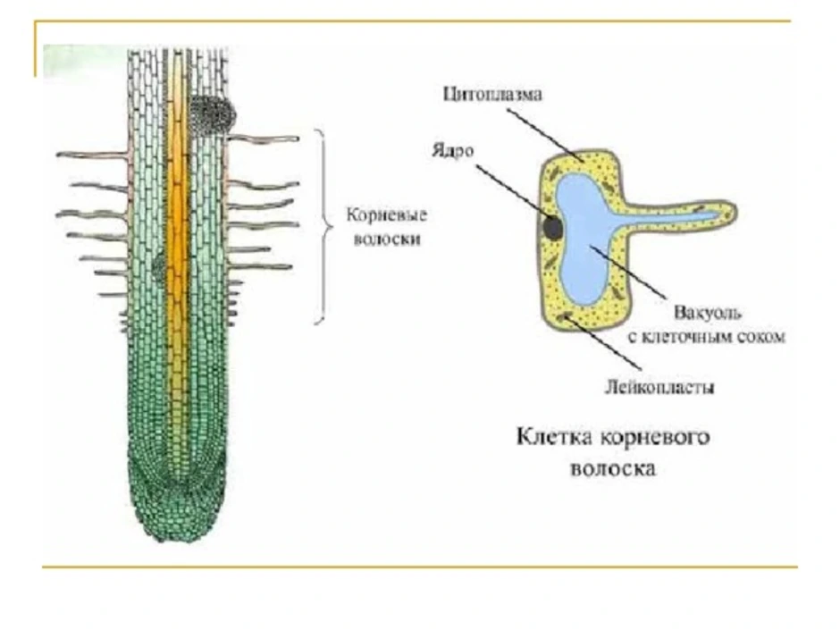
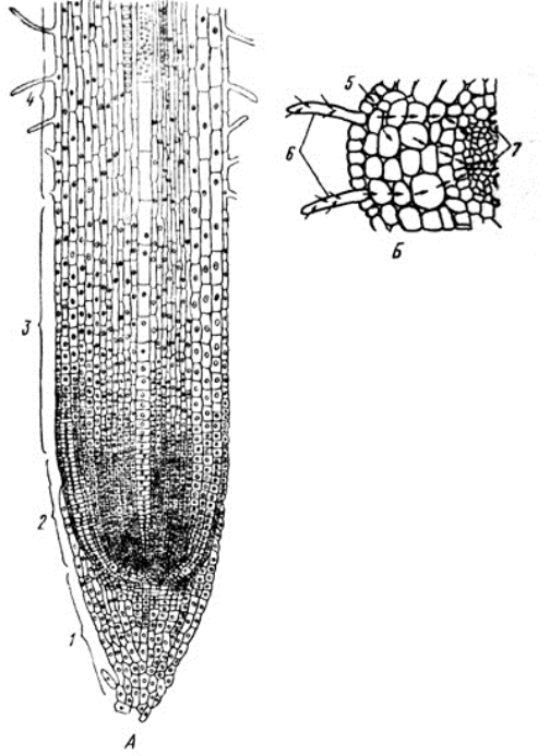
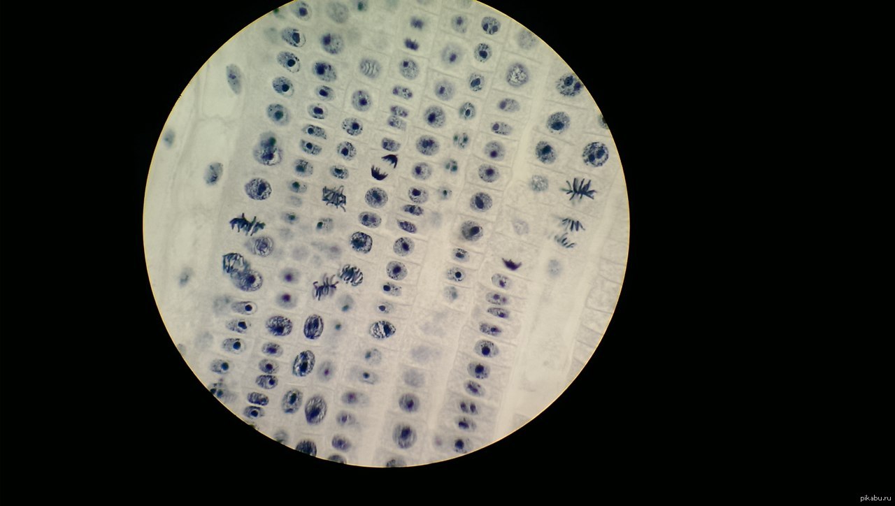
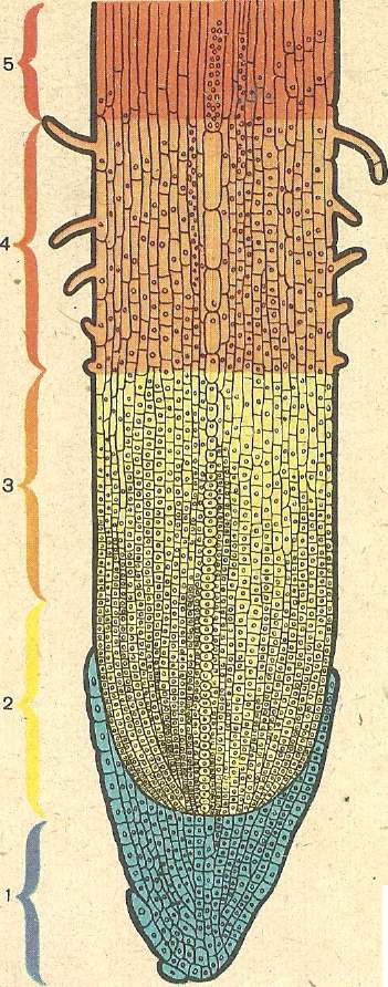
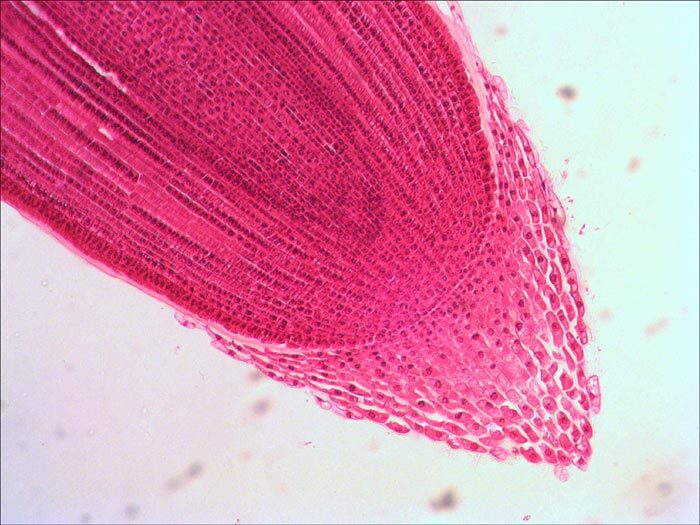
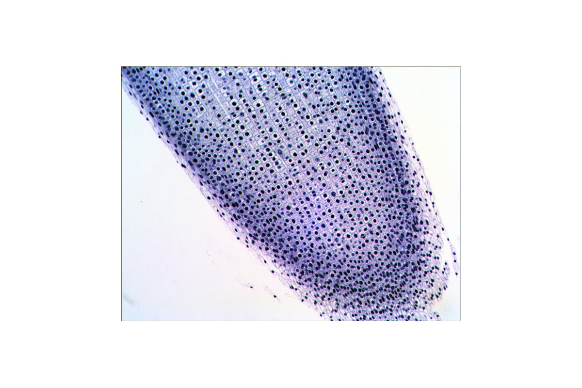
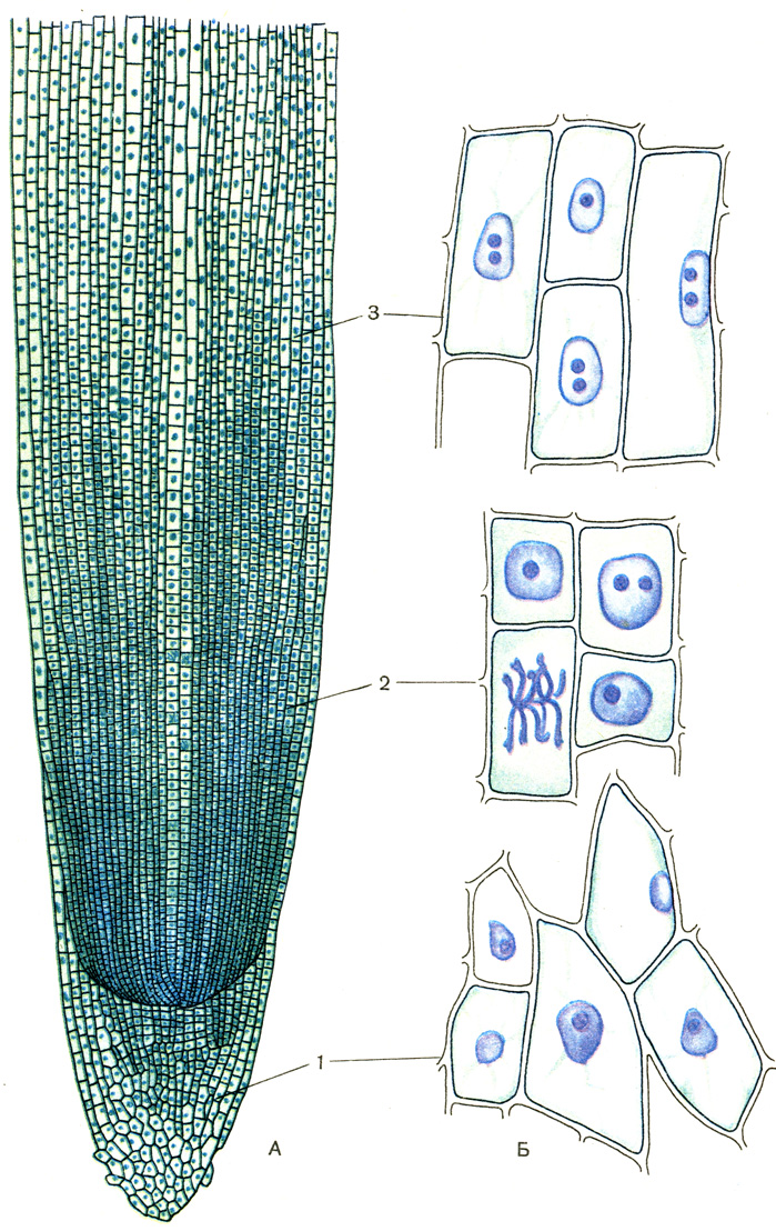
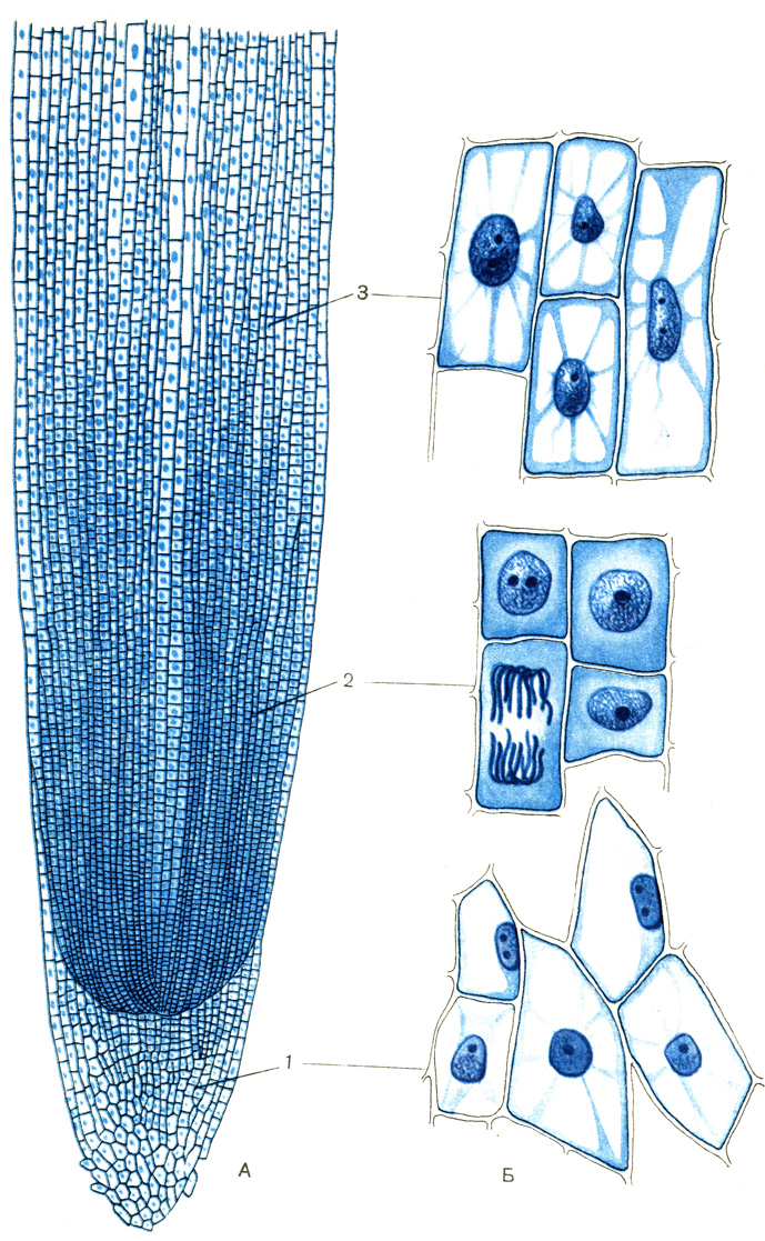
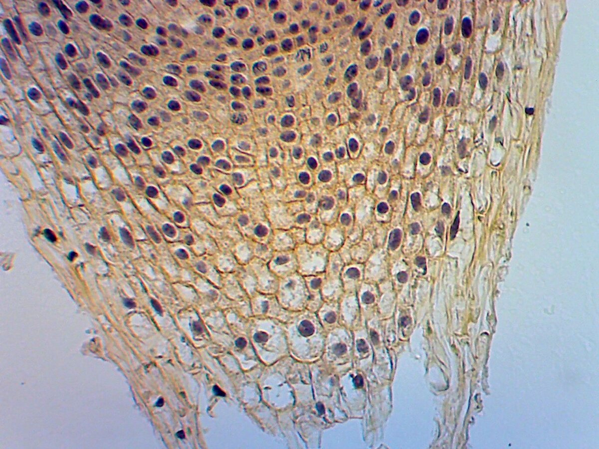
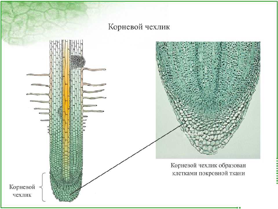
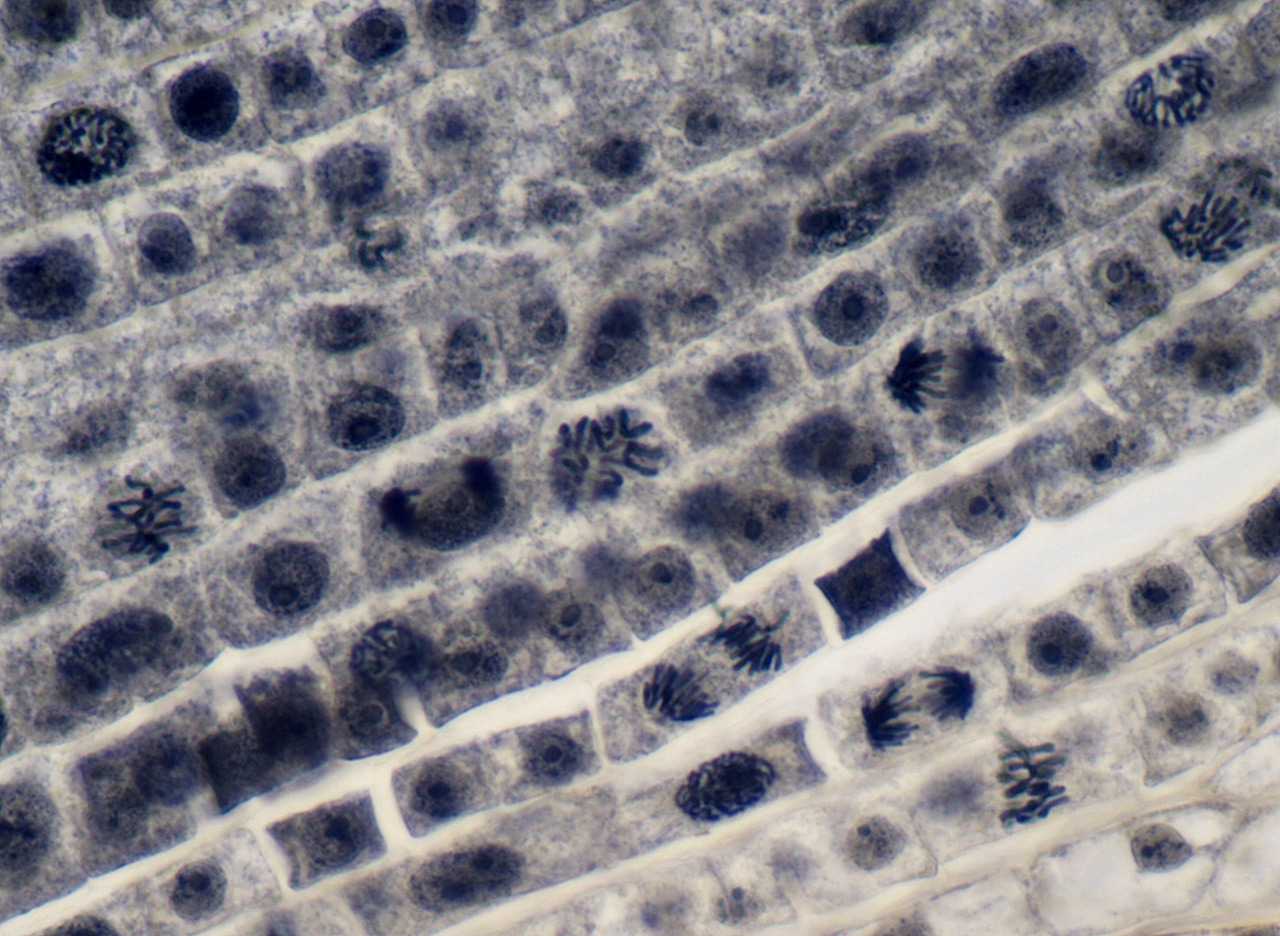
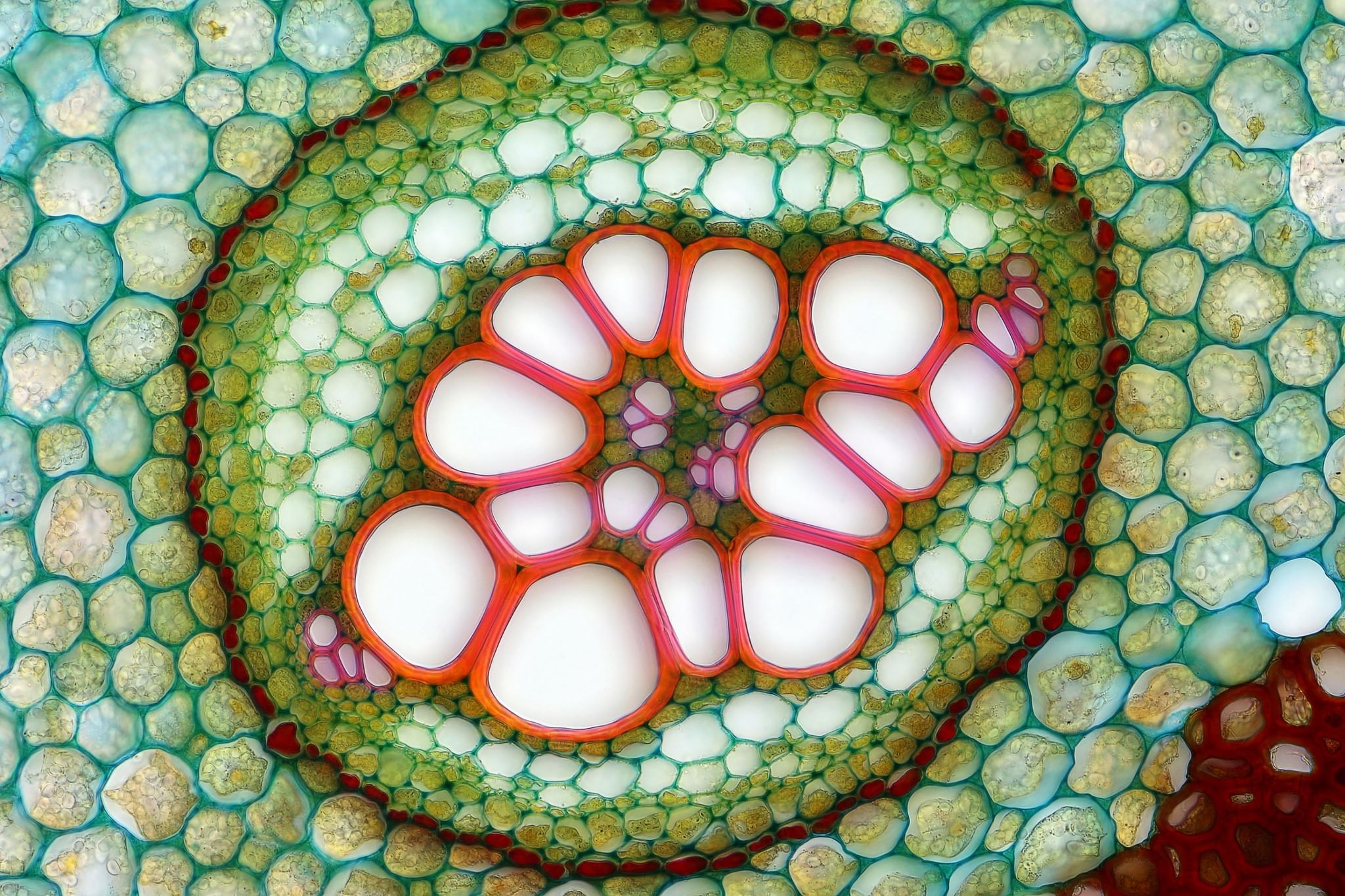
Microscopic photography, Microscopic, Plant cell imagesMitosis Cell in the Root Tip of Onion Under a Microscope. Stock Image - Image ofMitosis, Cell cycle, Fabric patternsOnion Root ImagesOnion Root ImagesOnion Root Tip Mitosis Labeled submited images Pic2Fly Mitosis, Teaching sciencePin by Alice Ham on Gene Visualisation Mitosis, Biology lessons, Cell biologyPlant gravity Pattern, Plants, DecorPlant Mitosis Onion Root Tip Stock Photo Royalty-Free FreeImagesRoot Tip of Onion and Mitosis Cell in the Root Tip of Onion Under a Microscope. Root Tip of Onion and Mitosis Cell in the Root Tip of Onion Under a Microscope. Root Tip: Root Apical Meristem, Protoderm, Procambium, Ground Meristem Biology, Root: the structure of the root. Types of roots (biology)Similax root bundles of a ranunculus. So beautiful! Patterns in nature, Things uTop Tips for Observing Mitosis Lab Mitosis, Meiosis, Meiosis activityTransverse section of part of a root of a monocot: Maize (Zea mays) Microscopic Word bank -signaling molecule -cell receptor -signal transduction pathway -nucleАнатомия корня 2020 ВКонтактеВ световой микроскоп можно увидеть органоиды: найдено 87 картинокВидеоокуляр ORBITOR 0,3 MPix (арт. 10474) купить в МосквеВПР по биологии 2019 - 5 класс - вариант 16Галерея фотографий. Что можно увидеть в микроскоп Levenhuk 630Галерея фотографий. Что можно увидеть в микроскоп Levenhuk 630Зона роста корневого чехликаЗоны кончика корня под микроскопом: найдено 87 изображенийКартинки КЛЕТКИ ЗОНЫ ВСАСЫВАНИЯ ИМЕЮТКарточки по биологииКарточки по биологииКлетках кончика корня: найдено 77 изображенийКорешок лука под микроскопом. ПикабуМатериалы на урокМеристемы. Функции и классификация меристем. Клеточное строение апекса и кончикаМикроскоп Discovery Pico с книгой(Цифровой), Цифровой, 400 крат купить по выгоднМитоз в корешке лука. Биологическое значение митоза состоит в строго одинаковом Набор микропрепаратов N20 NG Levenhuk 29277 - выгодная цена, отзывы, характеристРассмотрите рисунок растительной клетки какая структура клетки обозначена на рисРеакция на белки по Мэзиа 1969 Паламарчук И.А., Веселова Т.Д. - Изучение раститеРеакция на белки по Мэзиа 1969 Паламарчук И.А., Веселова Т.Д. - Изучение раститеСкачать обои природа, ветка, разрез, сосна, микроскоп, раздел макро в разрешенииСможешь угадать фото предметов под микроскопом? Профессор Гуглов ДзенСрезы органов растенийСтроение корня Ткань образующая корневой чехликФайл:Meristemo apical 1.jpg - ВікіпедіяФайл:Root tip.JPG - Википедия