Как выглядит нейрон под микроскопом - найдено 59 изображений
Найдено изображений: 59
Как выглядит нейрон под микроскопом2-Photon fluorescence image of glial cells in the cerebellum 2010 Photomicrograp
20 Dazzling Photos Of A Bizarre World You Need A Microscope To See HuffPost Micr30 Images Of Life Under A Microscope Things under a microscope, Microscopic photA scanning electron microscope (SEM) image zooms in on the baroque branching strAlexey Kashpersky on Behance #neuron #synapse #neural #brain Medical illustratioAstrocytic glial cell with cortical neuron, SEM - Stock Image - C036/9804 - ScieBook Review: Connectome Glial cells, Microscopic photography, Medical illustrati
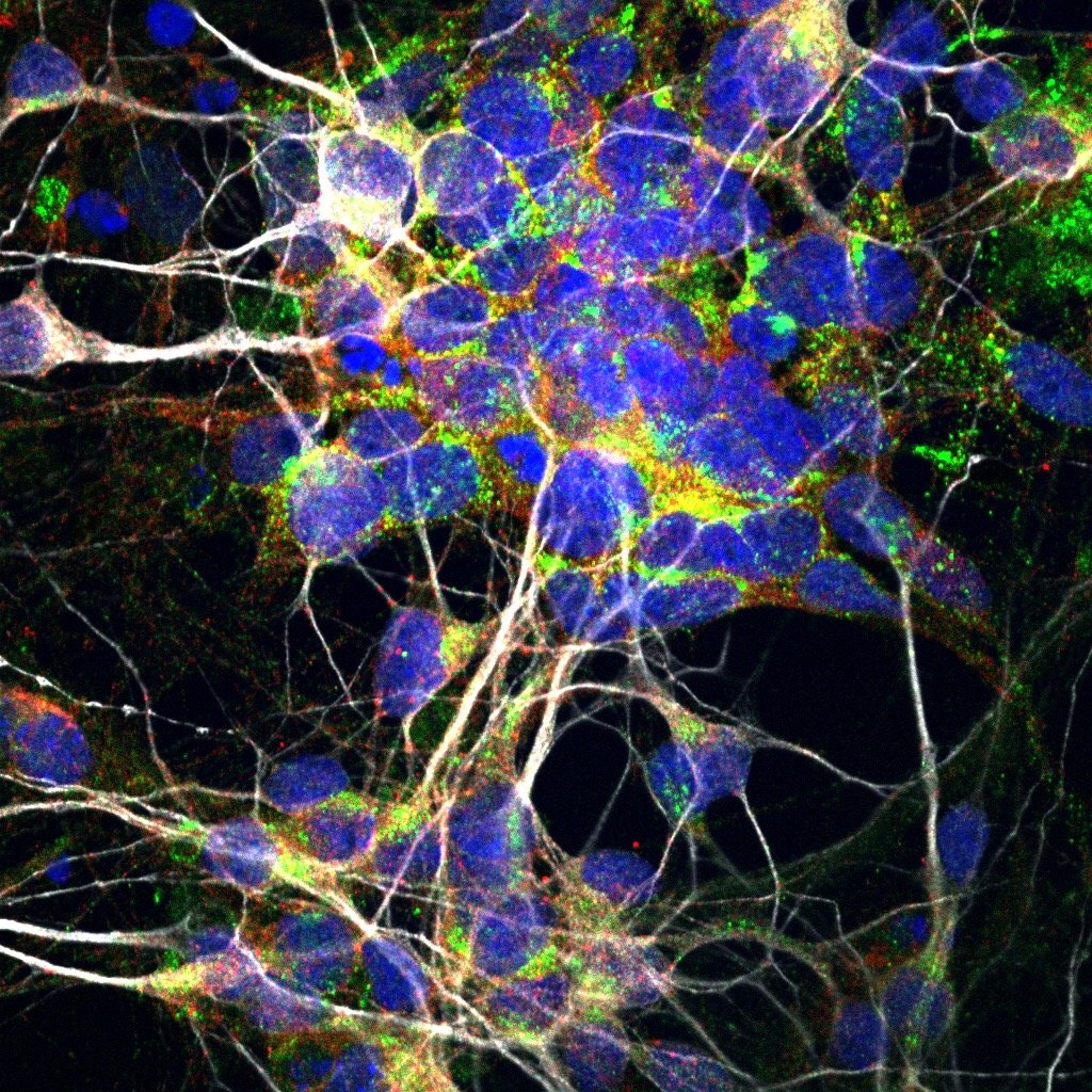
Brain cells, fluorescence micrograph - Stock Image C023/4113 Neuroscience art, MColored SEM of an oligodendrocyte. This cell forms the myelin sheaths around nerConfocal microscopy of mouse brain, cortex Confocal microscopy, Brain neurons, Ndesigur bilet Medicament neuron cell microscope aluminiu Nefavorabil naţionalismDiário Gottogreyoffwaffle, 45 anos de idade - Mamba - site gratuito de bate, стрDifferent Types of Nervous Tissue Microscopic photography, Nerve cell, Glial celExplore the Intricacies of a Neuron in the Hippocampus
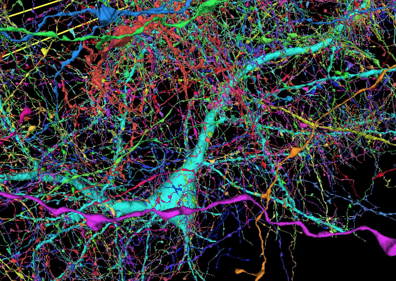
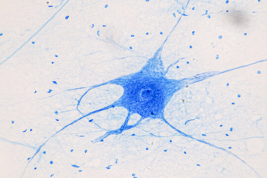
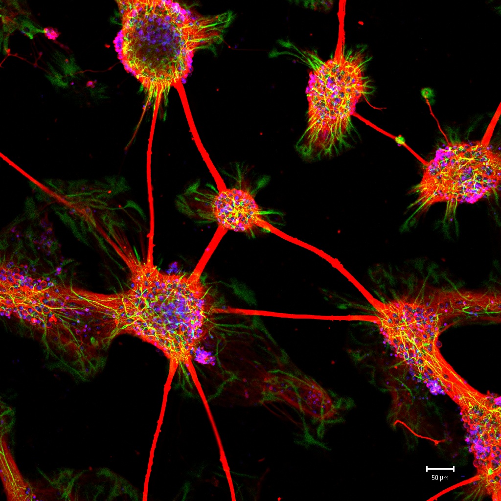
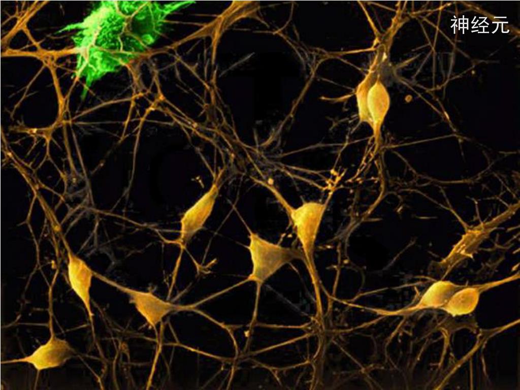
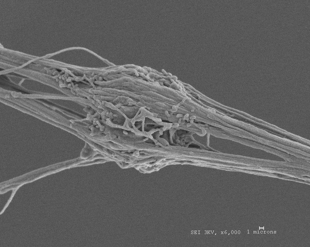
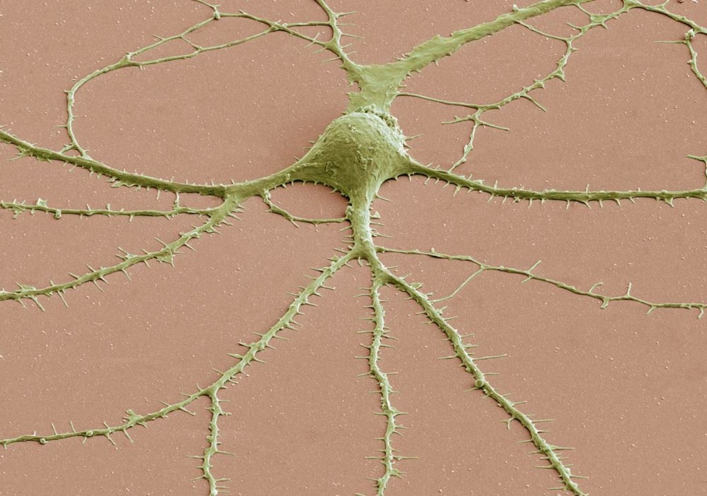
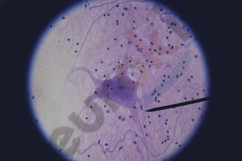
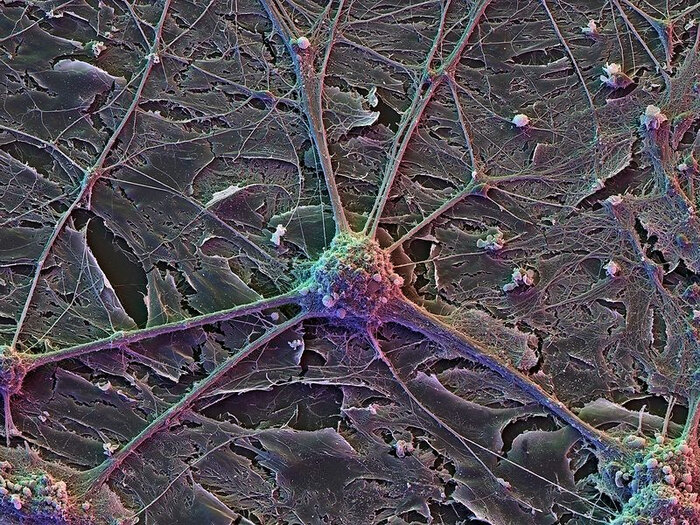
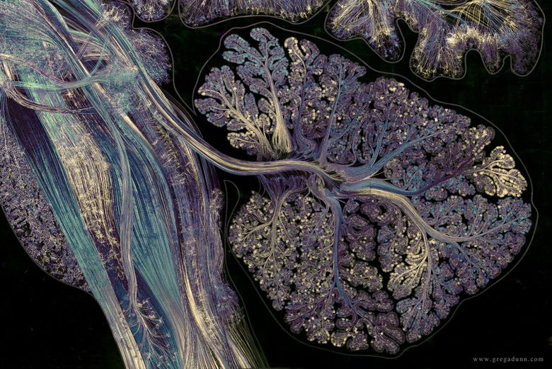
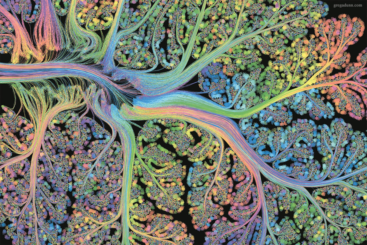
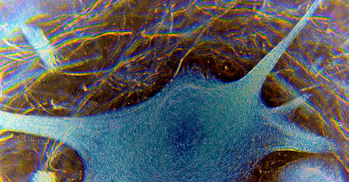
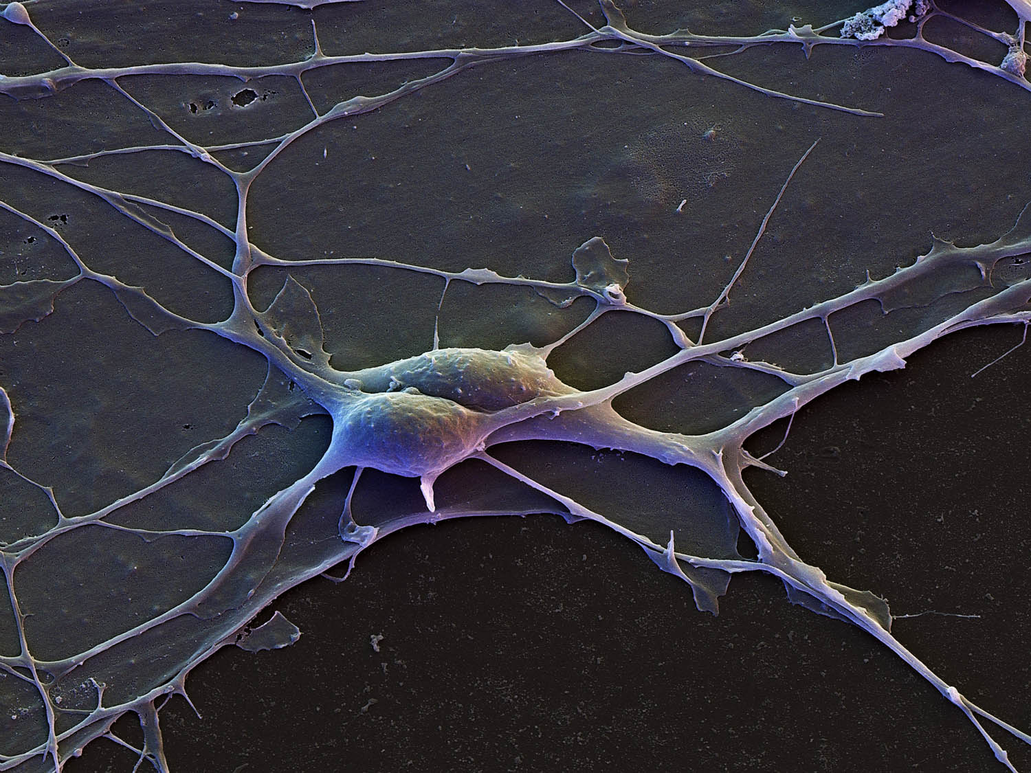
False-colour SEM of neurones from cerebral cortex - Stock Image - P360/0042 - ScFotografías tomadas por un microscopio electrónico que nos permite ver ... neuroHarvard and Google researchers have engineered a new 3D map of the human brain SHistology Изображения: просматривайте стоковые фотографии, векторные изображенияHistoric drawing of neurons Connectome: How the Brain's Wiring Makes Us Who We AHuman cerebral cortex neurons. LM X75 Neurons, Macro photography, Cerebral corteiLab Organizer :: Light Microscopy CoreImmune response in the human brain accurately measured for the first time ever. medicine- scientific photopraphy- scanning electron microscopy - inner organs, bmicroscopy on Tumblr Microscopic photography, Nerve cell, Science natureNerve Cells Microscopic photography, Brain art, Nerve cellNerve support cell (With images) Microscopic photography, Microscopic, MicroscopNeuron Art Neurons, Microscopic photography, Brain artNeuronally differentiated P19 cells 2009 Photomicrography Competition Nikon’s SmNeuroscience Gallery \ ConnCAD.com Micro photography, Science and nature, NeurosNew Brain Cells May Knock Out Old Memories Neurons, Memories, CellNikon Small World Photomicrography Competition Nikon small world, Microscopic phPin on Mad ScientistsPin on Work InspoPPT - 神 经 调 节 PowerPoint Presentation, free download - ID:5959042Purkinje nerve cell in the brain Electron microscope, Microscopic photography, NQuadruple fluorescence image revealing the complexity of the optic fiber layer oSingh Center for NanotechnologySmall World Competition 2004 Neurons, Differentiation, Small worldSpeed of brain-cell chatter clocked for first timeStriking, cutting-edge scientific images now on display at Washington Dulles IntThe 30,000 futures of the brain Nerve cell, Stem cells, NeuronsThe Big Picture: The hidden beauties unlocked by photomicrographs in 2024 Nikon The nervous tissue is the source of communication throughout the body. Nervous cThe toxic relationship between ALS and frontotemporal dementiaUC San Diego Health Sciences Spinal nerve, Scanning electron micrograph, MicroscUnleash the Power of Your Brain with Cortex NeuronsГДЗ параграф 6 Биология 9 класс Пасечник ФГОС Гарантия хорошей оценки ✅Двигательный нейрон под микроскопом. Стоковое Изображение - изображение насчитывКлетки мозга под микроскопом Пикабу ДзенКрасиво нарисованный синапс 2017 Анатомия человека ВКонтактеМаксим Руссо: Молекулярный механизм биологических часов Микроскопическая фотограМашинное обучение и Нейронные сети " - сообщество Яндекс.КьюНейроискусство: зачем создают картины из нейронов мозга / HabrНейрон под микроскопом - Olphoto.ruОда мозжечку: для чего нужен и за что отвечает малый мозг TechInsider ДзенОдинокий нейрон ПикабуПриматы отличаются от других млекопитающих архитектурой нейроновСПИН (Казак Елена Александровна) / Стихи.ру