Как выглядит мозг под микроскопом - найдено 60 изображений
Найдено изображений: 60
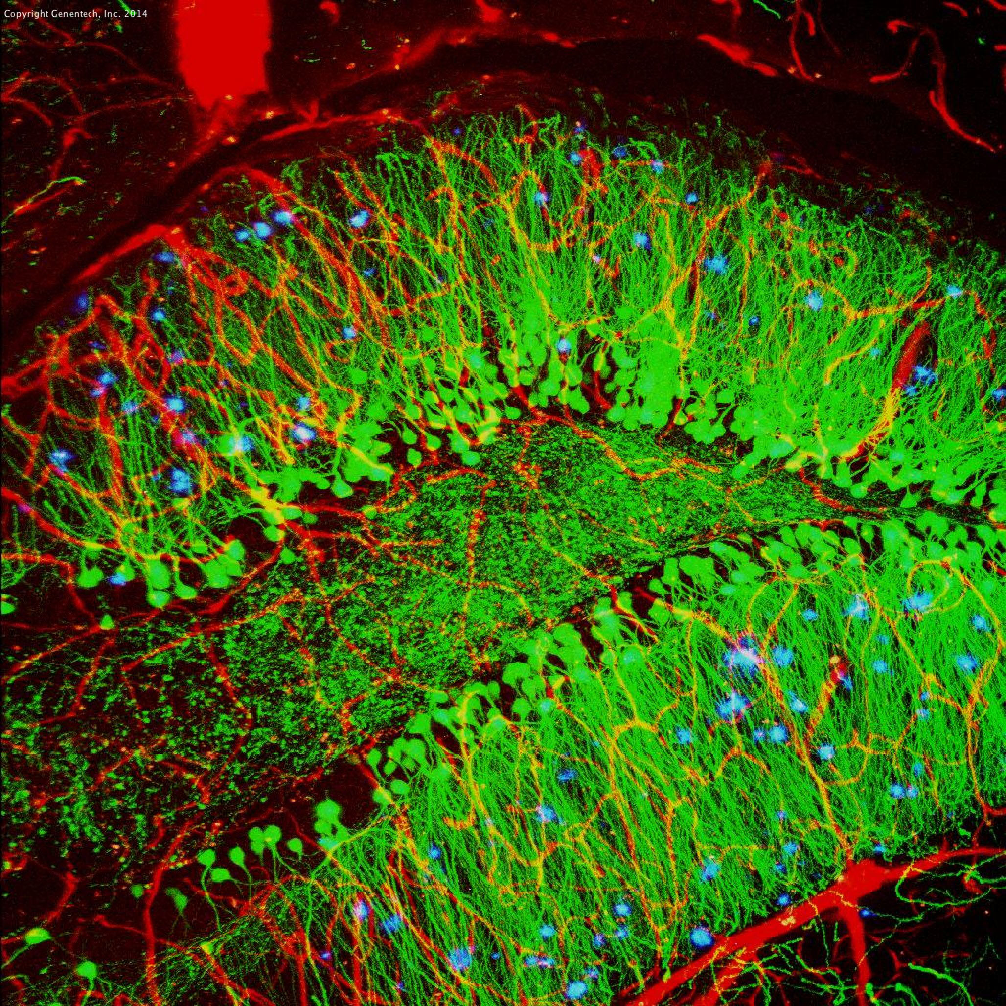
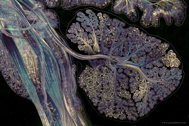
Как выглядит мозг под микроскопом2-Photon fluorescence image of glial cells in the cerebellum 2010 Photomicrograp
2007 Photomicrography Competition Nikon small world, Macro and micro, Small worl25 Stunning Microscopic Images That Show Hidden Universes In Everyday Objects Mi30+ Фантастических Фотографий Предметов И Существ Под Микроскопом Things under a447573.jpg Visuals Unlimited Neurons, Cerebral cortex, Chocolate cookieAlexey Kashpersky on Behance #neuron #synapse #neural #brain Medical illustratioAnt face, looking through microscope
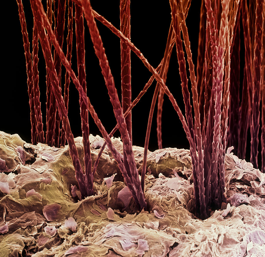
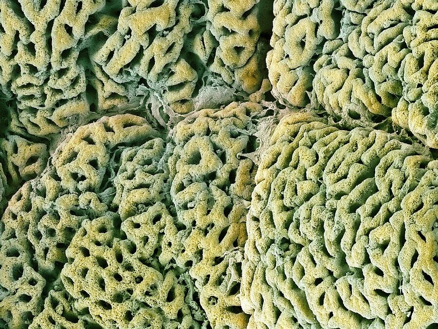
Beautiful Microscopic Images Of Fly Brains, Crustacean Claws, And Slime MicroscoBrain cells, fluorescence micrograph - Stock Image C023/4113 Neuroscience art, MCaterpillar: Things under a microscope, Microscopic photography, MicroscopicChoroid Plexus Secretory Cells, SEM' Photographic Print - Steve Gschmeissner AllConfocal microscopy of mouse brain, cortex Confocal microscopy, Brain neurons, NDatei:Dry blood under a microscope 4 (cropped).jpg - WikipediaDiário Gottogreyoffwaffle, 45 anos de idade - Mamba - site gratuito de bate, стр
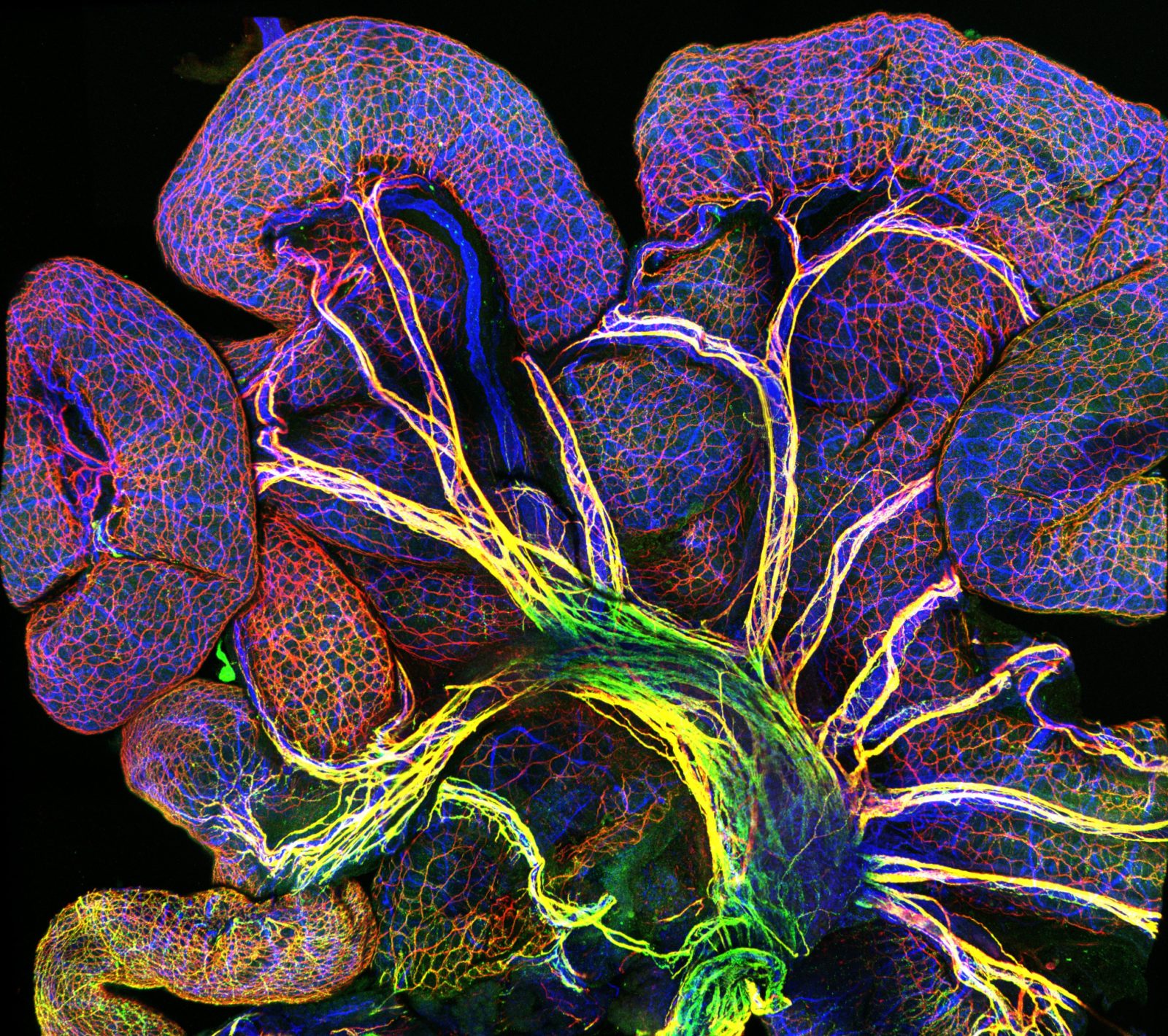
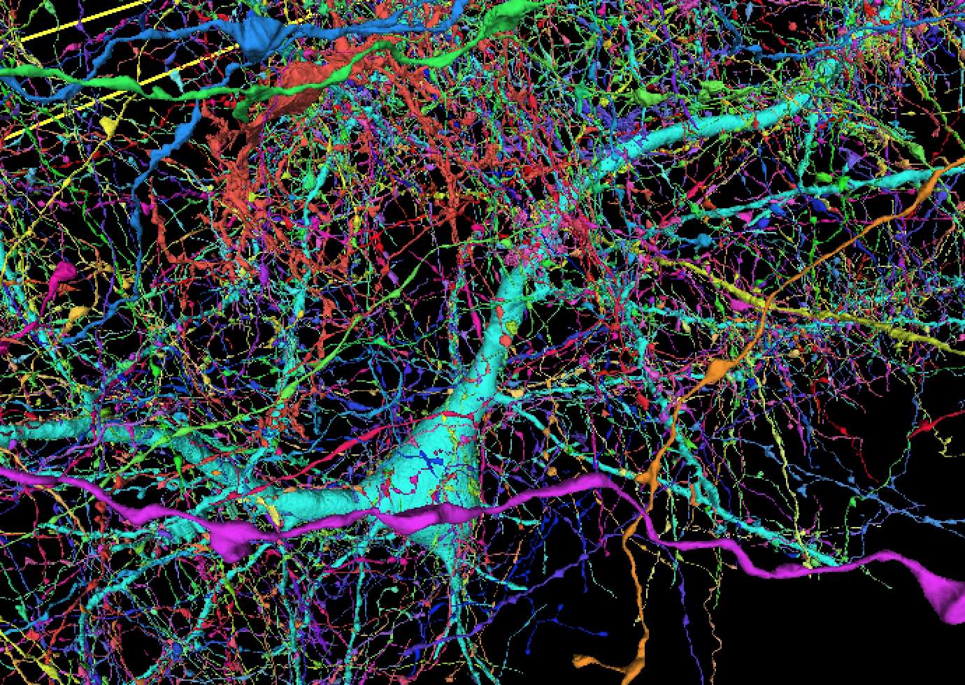
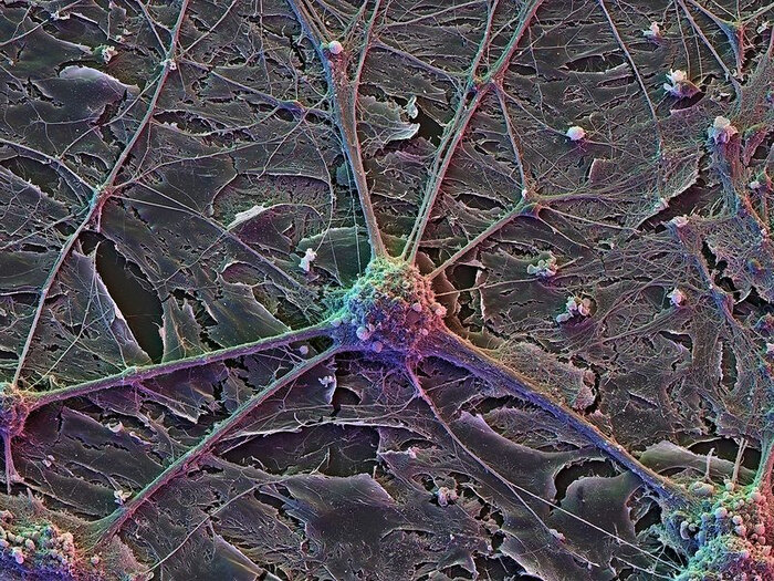
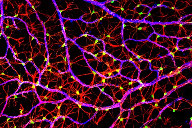
Different Types of Nervous Tissue Microscopic photography, Nerve cell, Glial celDog Hair, Sem #2 Photograph by Science Photo Library - Fine Art AmericaDr Lindsey Fitzharris (@DrLindseyFitz) on X Human body, Brain art, Human brainEndothelial cells in intestine of an 18.5-day old mouse embryo Nikon’s Small WorEndothelial cells in intestine of an 18.5-day old mouse embryo Nikon’s Small WorEpitelio ciliado vesicular False-colour SEM of neurones from cerebral cortex - Stock Image - P360/0042 - ScFile:Glomerulum of mouse kidney in Scanning Electron Microscope, magnification 1Fotografías tomadas por un microscopio electrónico que nos permite ver ... neuroGlial Cells, SEM' Photographic Print - Thomas Deerinck Art.com Fotografía microsHarvard and Google researchers have engineered a new 3D map of the human brain SHealthy Mouth, Healthy Body National Institute of Dental and Craniofacial Researmedicine- scientific photopraphy- scanning electron microscopy - inner organs, bMind-Blowing Brain Images From Then and Now Scanning electron micrograph, ElectrNerve Cells Microscopic photography, Brain art, Nerve cellNeuron Art Neurons, Microscopic photography, Brain artNeuronally differentiated P19 cells 2009 Photomicrography Competition Systems biNeuroscience Gallery \ ConnCAD.com Micro photography, Science and nature, NeurosPeering into the micro world Brain images, Vagus nerve, BrainPes Hipocampi Major Santiago Ramon Y Cajal by Science Source in 2020 MicroscopicPin by Leonie Hadorn on Uff in 2023 Neurons, Microscopic photography, Brain imagPin on Guardado rápidoPin on Microorganisms - MicroorganismoPurkinje nerve cell in the brain Electron microscope, Microscopic photography, NQuadruple fluorescence image revealing the complexity of the optic fiber layer oRagworm Mouth, Sem by Steve Gschmeissner Microscopic photography, Macro photograShort Sharp Science: Schizophrenic brain cells created in the labSmall World Competition 2004 Neurons, Nerve cell, Systems biologyStriking, cutting-edge scientific images now on display at Washington Dulles IntThe Big Picture: The hidden beauties unlocked by photomicrographs in 2024 Nikon This illustration shows a synapse. When an action potential arrives at a synapseThis will bug you... The indestructible micro-animals that can survive the vacuuTop images from the 2014 Nikon Small World Competition Small world, Nikon, PhotoUC San Diego Health Sciences Spinal nerve, Scanning electron micrograph, MicroscЖизнь под микроскопом: срез спинного мозгаКак выглядит человеческое тело под микроскопом Профессор Гуглов ДзенКак выглядят наши органы под микроскопомКлетки мозга под микроскопом Пикабу ДзенКонфокальная микроскопия - azimp-micro.ruМашинное обучение и Нейронные сети " - сообщество Яндекс.КьюМикрофотография поперечного сечения нервного пучка. - Мы увеличиваем вещи как ниНейроискусство: зачем создают картины из нейронов мозга / HabrСамые ужасные микроскопические монстры Microscopic images, Scanning electron micСкачать 1400x1050 нервные клетки, сплетение, 3d обои, картинки стандарт 4:3Учёные приблизились к победе над эпилепсией, шизофренией и склерозом